Marek Chorazewicz's Photo Gallery
VHU-C12Murphyite Pb(Te6+O4)

Field of View: 400 μm
Largest Crystal Size: 25 μm
Clear prismatic crystals of murphyite. Other areas on the same specimen have the following confirmed minerals: ottoite, shieffelinite, emmonsite, and "girdite," a discredited mineral, likely a mixture of ottoite and plumbotellurite.
Please see the parent photo for more details.
The photo was taken by Michael Cox on his Keyence microscope at the Pacific Micro Mineral Conference, Fallbrook, Jan 2024.
VHU-C12Murphyite Pb(Te6+O4)
Field of View: 400 μm
Largest Crystal Size: 40 μm
Clear prismatic crystals of murphyite. Other areas on the same specimen have the following confirmed minerals: ottoite, shieffelinite, emmonsite, and "girdite," a discredited mineral, likely a mixture of ottoite and plumbotellurite.
Please see the parent photo for more details.
The photo was taken by Michael Cox on his Keyence microscope at the Pacific Micro Mineral Conference, Fallbrook, Jan 2024.
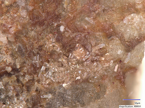
VHU-C12Murphyite Pb(Te6+O4) , Ottoite Pb2TeO5

Field of View: 350 μm
Largest Crystal Size: 50 mm
Clear prismatic crystals of murphyite. Other areas on the same specimen have the following confirmed minerals: ottoite, shieffelinite, emmonsite, and "girdite," a discredited mineral, likely a mixture of ottoite and plumbotellurite.
The murphyite crystals have quite similar morphology to raspite. The tellurium-rich raspite from Tombstone has been noted and investigated a decade ago. However, more recently, Bruce Murphy found clear crystals on a specimen from Sid Williams and sent them to the University of Arizona. They measured a higher Te/W ratio, and that's how murphyite was described.
On this particular specimen, the molecular ratio of Te/W is close to 50/50%, but the definitive identification came from the single crystal XRD spectrum taken at the Australian Synchrotron of Monash University, thanks to Stuart Mills -- it was a match for murphyite. Bruce Murphy was very happy to learn that more of this mineral had been found. Unfortunately, it was just before his death.
The photo was taken by Michael Cox on his Keyence microscope at the Pacific Micro Mineral Conference, Fallbrook, Jan 2024.
Dr. Matthias Weil

The mineral matthiasweilite is named after Dr. Weil.
NFX-63DMatthiasweilite PbTe4+O3 , Northstarite Pb6(Te4+O3)5(S6+O3S2-)

Field of View: 0.68 mm
Matthiasweilite (light yellow) partially filling a cavity in quartz. The darker yellow crystals on the left are northstarite. The field of view is 0.68 mm across. Type specimen - Natural History Museum of Los Angeles County catalogue number 76156.
Photo taken by Dr. Anthony Kampf on Keyence digital scope at NHMLAC.
1VT-053Backite Pb2AlTeO6Cl
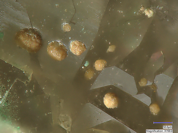
Field of View: 500 μm
Closeup on sparkly tan backite balls on quartz. Some reflections from tiny backite crystals can be seen at the edges of the hemispheres. In the lower-left corner, there is a small fuettererite incursion. See the parent photo for more info.
Identified by Dr. Housley with Raman spectroscopy at Caltech Geoplanetary lab in Pasadena, CA. Michael Cox took the picture on Keyence digital microscope at Pacific Micro Mineral Conference, Jan 2023.
1VT-053Fuettererite Pb3Cu2+6Te6+O6(OH)7Cl5 , Backite Pb2AlTeO6Cl

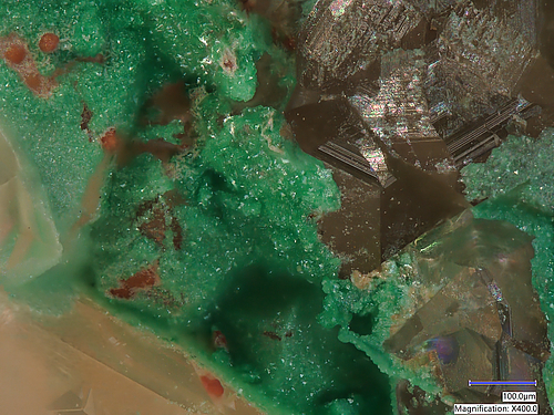
Field of View: 750 μm
Red backite balls with blue-green fuettererite on quartz crystals. This is the first confirmed find of these two extremely rare hexavalent tellurium oxysalts minerals from the Aga mine. They have been found at other Otto Mtn localities but not there. Associated with yellow-green bairdite platelets, tan backite balls, and green-blue paratacamite crystals, not shown here.
Identified with Raman spectroscopy at Caltech Geoplanetary lab in Pasadena, CA. The picture was taken by Michael Cox on Keyence digital microscope at Pacific Micro Mineral Conference, Jan 2023.
Sulfur Hole area

In 2019 the Sulfur Hole was completely covered by debris from the rock layers higher up. Looks more like a mound than a hole. However, some good sulfates have been found from the hydrothermal chimney directly above the hole location, including the first arsenate from the locality, scorodite.
C2Y-YMEScorodite Fe3+AsO4·2H2O , Copiapite Fe2+Fe3+4(SO4)6(OH)2·20H2O , Metavoltine K2Na6Fe2+Fe3+6O2(SO4)12·18H2O

Field of View: 5 mm
This material is from the first verified find of arsenate mineral at the Sulfur Hole by MarekC and Bob Housley.
Collected from the side wall of the hydrothermal chimney directly above the hole during April 2019 MSSC post-fieldtrip to Borate area. Raman verified.
Cellphone photo thru the eyepiece of the microscope.
WCV-N1YCopiapite Fe2+Fe3+4(SO4)6(OH)2·20H2O , Sulphur S8 , Jarosite KFe3+3(SO4)2(OH)6

Field of View: 14 mm
Collected from the side wall of the hydrothermal chimney directly above the hole during April 2019 MSSC post-fieldtrip to Borate area. Raman verified.
Cellphone photo thru the eyepiece of the microscope.


